About MRI of the Spine and Brain
What is magnetic resonance imaging (MRI)?
Magnetic resonance imaging (MRI) uses a large magnet, radiofrequencies, and a computer to produce detailed images of organs and structures within the body, in this case, the brain and spine. MRI is used to help diagnose a health problem.
The MRI machine is a large, tube-shaped machine that creates a strong magnetic field around the patient. Some look like narrow tunnels. Others are more open. This magnetic field, along with a radiofrequency, alters the hydrogen atoms’ natural alignment in the body. Computers are then used to form two-dimensional (2D) images of the brain and/or spine based on the activity of the hydrogen atoms. Cross-sectional views can be done to show more details. MRI does not use radiation, like X-rays or computed tomography (CT scans).
Magnetic resonance (MRI) may be used instead of computed tomography (CT) when organs or soft tissue are being studied. This is because with MRI scanning bones do not obscure the images of organs and soft tissues, as does CT scanning.
Functional magnetic resonance imaging of the brain (fMRI) is used to determine the specific location of the brain where a certain function, such as speech or memory, happens. The general areas of the brain in which such functions happen are known, but the exact location may vary from person to person. During fMRI imaging of the brain, you will be asked to do a specific task, such as recite the Pledge of Allegiance, while the scan is being done. By pinpointing the exact location of the functional center in the brain, healthcare providers can plan surgery or other treatments for certain brain disorders.
Why might I need an MRI?
MRI may be used to check the brain and/or spinal cord for injuries, the presence of structural abnormalities, or certain other conditions, such as:
- Tumors
- Abscesses (collections of pus)
- Congenital defects (those you are born with) of the spine
- Aneurysms (weakening and ballooning of an artery)
- Venous malformations (abnormal and dilated veins)
- Bleeding into the brain or spinal cord
- Subdural hematoma (an area of bleeding just under the dura mater, or covering of the brain)
- Degenerative diseases, such as multiple sclerosis, hypoxic encephalopathy (dysfunction of the brain due to a lack of oxygen), or encephalomyelitis (inflammation or infection of the brain)
- Hydrocephalus (fluid in the brain)
- Herniation or degeneration of discs of the spine
- To help plan surgeries on the spine, such as decompression of a pinched nerve or spinal fusion
- To look for problems after surgery, such as scarring or infection
MRI can also help to identify the specific part of the brain controlling a function, such as speech or memory, to assist in treatment of a condition of the brain.
There may be other reasons for your healthcare provider to recommend MRI of the spine or brain.
What are the risks of an MRI?
There is no risk of exposure to radiation during an MRI procedure.
Due to the use of the strong magnet, MRI cannot be used for people with the following:
- Implanted pacemakers
- Some older intracranial aneurysm clips
- Cochlear implants
- Certain prosthetic devices
- Implanted drug infusion pumps
- Neurostimulators
- Bone-growth stimulators
- Certain intrauterine contraceptive devices
- Any other type of iron-based metal implants
- Internal metallic objects such as bullets or shrapnel, surgical clips, pins, plates, screws, metal sutures, or wire mesh
If you are pregnant or think you may be, tell your healthcare provider. In general, there is no known risk of MRI in pregnancy. However, particularly in the first trimester, MRI should only be used to address very important problems or suspected abnormalities.
If contrast dye is used, there is a risk for allergic reaction to the dye. If you are allergic to or sensitive to medicines, contrast dye, or iodine, tell your healthcare providers.
MRI contrast may have an effect on other conditions. These include allergies, asthma, anemia, low blood pressure, kidney disease, and sickle cell disease.
Nephrogenic systemic fibrosis (NSF) is a very rare but serious complication of MRI contrast use in people with kidney disease or kidney failure. If you have a history of kidney disease, kidney failure, kidney transplant, liver disease, or are on dialysis, be sure to tell the MRI technologist or radiologist before getting the contrast dye.
There may be other risks depending on your specific medical condition. Be certain your healthcare provider knows about all of your medical conditions.
How to prepare?
Your healthcare provider will explain the procedure to you and give you a chance to ask questions. Make a list of questions and discuss these and any concerns with your healthcare provider before the procedure. Consider bringing a family member or trusted friend to the medical appointment to help you remember your questions and concerns and to take notes.
If your procedure involves the use of contrast dye, you will be asked to sign a consent form that gives permission to do the procedure. Read the form carefully and ask questions if anything is not clear.
Generally, there is no special restriction on diet or activity prior to an MRI procedure.
Before the MRI, it is extremely important that you inform the technologist if any of the following apply to you:
- You are claustrophobic and think that you will be unable to lie still inside the scanning machine, in which case you may be given a sedative
- You have a pacemaker or have had heart valves replaced
- You have any type of implanted pump, such as an insulin pump
- You have metal plates, pins, metal implants, surgical staples, or aneurysm clips
- You have any metallic fragments anywhere in the body
- You have permanent eyeliner or tattoos
- You are pregnant or think you may be pregnant
- You have ever had a bullet wound
- You have ever worked with metal (for example, a metal grinder or welder)
- You have any body piercings
- You have an intrauterine device (IUD)
- You are wearing a medicine patch
There is a possibility that you may get a sedative before the procedure, so you should plan to have someone drive you home afterward.
Based on your medical condition, your healthcare provider may request other specific preparation.
What happens during an MRI?
MRI may be done on an outpatient basis or as part of your stay in a hospital. Procedures may vary depending on your condition and your healthcare provider’s practices.
Generally, MRI of the spine and brain follows this process:
- You will be asked to remove any clothing, jewelry, eyeglasses, hearing aids, hairpins, removable dental work, or other objects that may get in the way of the procedure.
- If you are asked to remove clothing, you will be given a gown to wear.
- If you are to have an MRI with contrast, an intravenous (IV) line will be started in your hand or arm for injection of the contrast dye.
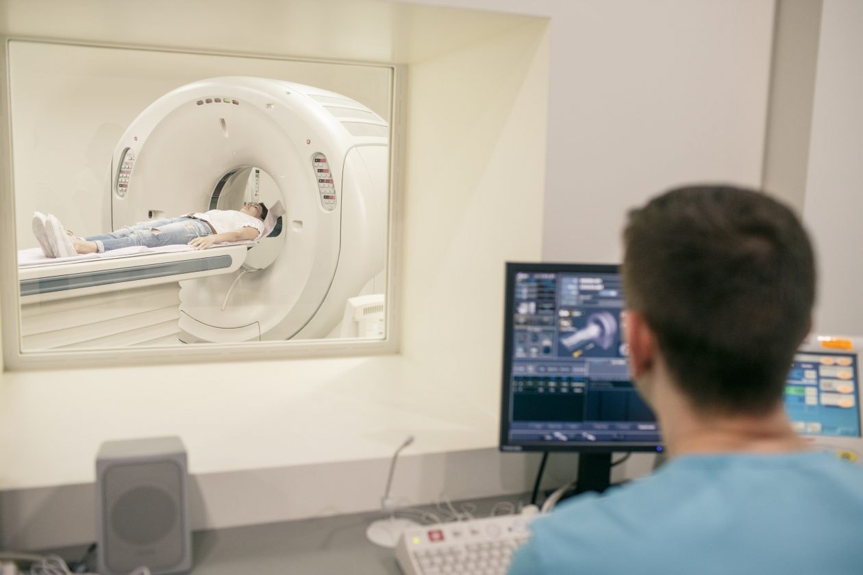
- You will lie on a narrow table that slides into the large circular opening of the scanning machine. Pillows and straps may be used to help prevent movement during the scan.
- The technologist will be in another room where the scanner controls are located. However, you will be in constant sight of the technologist through a window. Speakers inside the scanner allow technologist to talk to you and hear you. You will have a call button so that you can let the technologist know if you have any problems during the procedure. The technologist will be watching you at all times and will be in constant communication.
- You will be given earplugs or a headset to wear to help block out the noise from the scanner. Some headsets may provide music for you to listen to. During the scanning process, you will hear clicking and thumping noises as the magnetic field is created and pulses of radio waves are sent from the scanner.
- It will be important for you to stay very still during the exam. Any movement could cause distortion and affect the quality of the scan.
- At intervals, you may be told to hold your breath, or to not breathe for a few seconds. You will then be told when you can breathe. You should not have to hold your breath for longer than a few seconds.
- If contrast dye is used, you may feel some effects when the dye is injected into the IV line. These effects include a warm flushing sensation or a feeling of coldness, a salty or metallic taste in the mouth, a brief headache, itching, or nausea. These effects usually only last for a few moments.
- You should tell the technologist right away if you feel any breathing difficulties, sweating, numbness, or heart palpitations.
- Once the scan is done, the table will slide out of the scanner and you will be helped off the table.
- If an IV line was put in, it will be removed.
While the MRI itself causes no pain, having to lie still for the length of the procedure might cause some discomfort or pain, particularly if you’ve recently been injured or had surgery. The technologist will use all possible comfort measures and complete the procedure as quickly as possible to reduce any discomfort or pain.
On occasion, some people with metal fillings in their teeth may experience some slight tingling of the teeth during the procedure.
What happens after an MRI?
Move slowly when getting up from the scanner table to avoid any dizziness or lightheadedness from lying flat for the length of the procedure.
If any sedatives were used for the procedure, you may need to rest until the sedatives have worn off. You will also need someone to drive you home.
If contrast dye is used, you may be monitored for a period for any side effects or reactions to the contrast dye, such as itching, swelling, rash, or difficulty breathing.
If you notice any pain, redness, and/or swelling at the IV site after you go home, tell your healthcare provider as this could be a sign of infection or other type of reaction.
Otherwise, there is no special type of care needed after a MRI scan of the spine and brain. You may go back to your usual diet and activities, unless your healthcare provider tells you differently.
Your healthcare provider may give you additional or alternate instructions after the procedure, depending on your particular situation.
Next steps
Before you agree to the test or the procedure make sure you know:
- The name of the test or procedure
- The reason you are having the test or procedure
- What results to expect and what they mean
- The risks and benefits of the test or procedure
- What the possible side effects or complications are
- When and where you are to have the test or procedure
- Who will do the test or procedure and what that person’s qualifications are
- What would happen if you did not have the test or procedure
- Any alternative tests or procedures to think about
- When and how will you get the results
- Who to call after the test or procedure if you have questions or problems
- How much will you have to pay for the test or procedure